In the diagrams represent the structures labeled by single Rab proteins, their relative abundance and their localization within the cell.
Rab-labeled structures in the cells
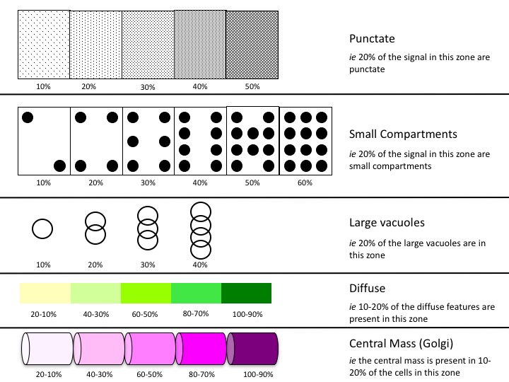
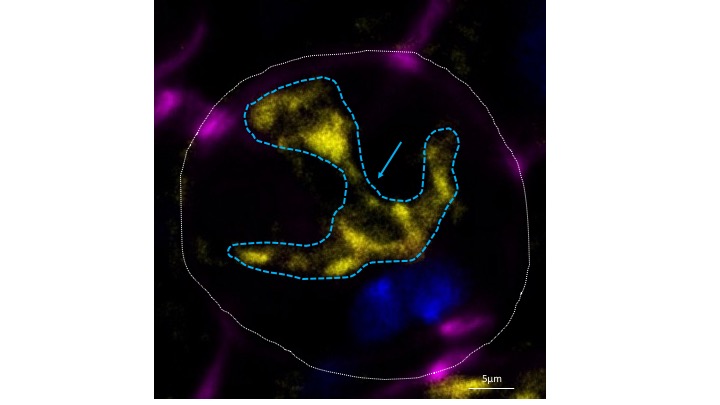
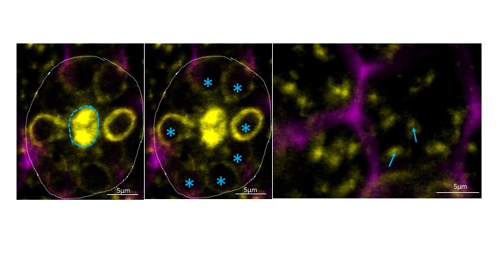
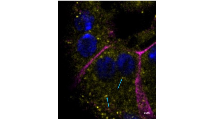
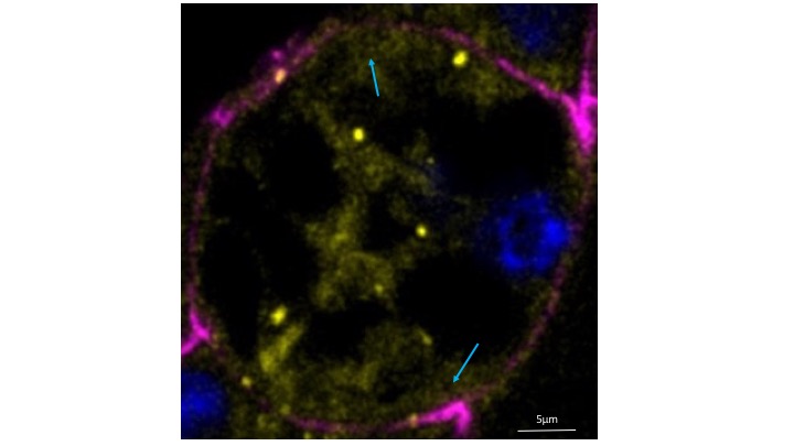
Different terms will be used to describe the Rab-marked structures found in the accessory gland. First, “central mass” which is a dense filamentous-like structure or central well, whose mass center is located at the center of the secondary cell (Figure 2 and 3A); “vacuoles” refer to hollow and membrane-delimited structures whose diameter can vary from 0.3μm to 8μm (Figure 3B). “small compartments” are features >0.5µm whose intra-cellular space is not visible (Figure 3C). “punctate” structures are particulate structures smaller than 0.5μm in diameter (Figure 4). And finally “diffuse” features describe staining that is spread out without visible particulate structures (Figure 5). On the summary diagrams, each of the structures is represented by a symbol according to the following key:
 |
| Figure 2 |
 |
| Figure 3A, 3B, 3C |
 |
| Figure 4 |
 |
| Figure 5 |
Location definitions
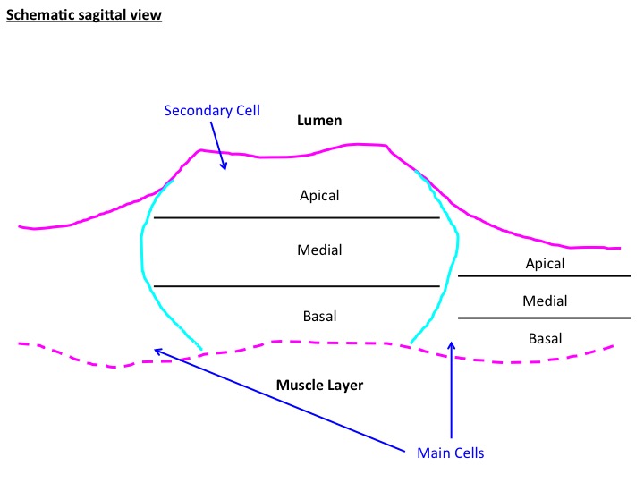
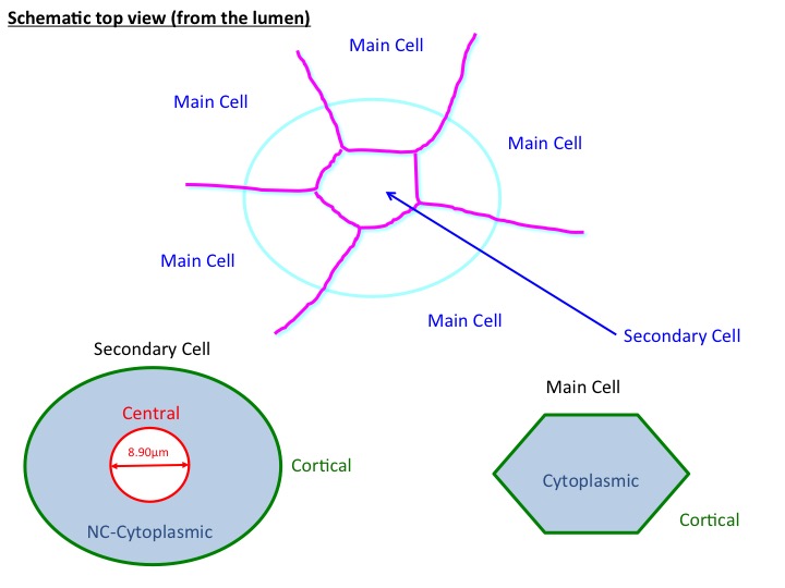
To describe the position of each of the Rab-labeled structures within the various cell types, we defined zones along the apical-basal axis and radial zones emanating from a line going through the cell’s center of mass along the apical-basal axis (see Figure 7 and Figure 8). To make the zones along the apical basal axis, we simply divided the cell into three, approximately equal zones (as defined by DE-CAD and DLG staining) called: apical, medial and basal. The radial zones were defined differently in each cell type. For the main cells, two zones were defined. Structures that touch the lateral plasma membrane were called “Cortical” and all other interior structures were simply called “Cytoplasmic”. For the secondary cells, three radial zones were defined. “Central” features correspond to those that are within an 8.90µm diameter cylinder centered on the central axis. “Cortical” features refer to those that are in contact with the lateral plasma membrane. Meanwhile all other structures are simply referred to as “non-central, cytoplasmic” or “NC cytoplasmic (Figure 8 ).
 |
| Figure 7 |
 |
| Figure 8 |